


Mr Frederick, a 35 year old construction worker presents with haematemesis. His endoscopy showed:
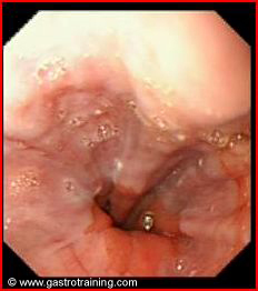
What is the diagnosis?
Oesophageal varices
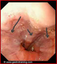
What are endoscopic stigmata of variceal bleed?
Red colour signs (cherry-red spots and red wale markings)-, are associated with higher risk of haemorrhage. These signs are thought to represent focal weakness in the variceal wall.
Fibrin clots or ‘white nipple sign’ are occasionally seen over the variceal columns which have recently bled.
Define small and large varices?
Varices are either small or large. Large varices (for the purpose of treatment) are those varices that are greater than 5 mm or tortuous veins or occupying more than one-third of the oesophageal lumen.
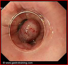
Discuss the endoscopic management of oesophageal varices?
Endoscopic variceal ligation (banding) is done to control variceal bleeding. The oesophageal mucosa and the submucosa containing varices are ensnared, causing subsequent strangulation, sloughing, and eventual fibrosis, resulting in obliteration of the varices.
Variceal band ligation therapy is superior to sclerotherapy in terms of rebleeding, all cause mortality and death due to bleeding in patients with bleeding oesophageal varices.
What are the endoscopic signs indicated increased risk of further bleeding?
The presence of variceal red colour signs (eg, cherry red spots, red whale markings, blue varices) indicates an increased risk of further bleeding.
Further reading

