


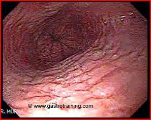
Mr Matthews, a 19 year old man presents with 2 year history of intermittent dysphagia. His endoscopy showed:
What is the diagnosis?
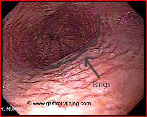
Eosinophilic oesophagitis. The picture shows a ringed appearance i.e. trachealized oesophagus.
Discuss the endoscopic findings in eosinophilic oesophagitis?
Endoscopic findings are usually subtle and a careful examination is needed along with biopsies. Endoscopic findings include:
- Strictures
- Trachealized oesophagus (ringed appearance)
- Whitish elevated papules that resemble candidiasis
- Longitudinal linear furrows (also called oesophageal corrugation)
How do you establish the diagnosis?
At least 5 biopsies should be obtained from the proximal and distal third of the oesophagus for optimal sensitivity.
The current accepted number of eosinophils needed for diagnosis is 15 eosinophils/HPF in the presence of a consistent clinical context.
Oesophageal biopsies should be obtained if the clinical history is suggestive (even if the oesophagus looks normal endoscopically)
What is Eosinophilic oesophagitis?
- Chronic oesophageal inflammation of unknown origin that is characterized by dense infiltration of eosinophils.
- It has been described in patients of all ages, however it is most common in the childhood
- It predominantly affects males
- Possibly allergic in aetiology as majority of patients have a personal or family h/o allergy
- New disease with an increasing incidence
Discuss the clinical manifestations of EO?
- Intermittent dysphagia
- Food impaction
- GORD like symptoms unresponsive to PPI
- Vomiting
- Chest pain
Further reading
Link to Eosinophilic oesophagitis
Image courtesy of www.gastrointestinalatlas.com

