Localisation and lesion recognition at Colonoscopy
Localisation
- The ileo-caecal valve is the only definite anatomical landmark in the colon. It may occasionally be difficult to find
- The internal appearance of transverse colon is usually triangular. However the descending colon may look triangular or the transverse colon may look circular
- There may be bluish/grey indentation from the liver at the hepatic flexure; however a similar appearance may sometimes occur at the splenic flexure.
- The distance of the lesion should only be mentioned on withdrawal. So you could say that the lesion/polyp was at 30cms in the sigmoid colon on withdrawal. The scope distance information at insertion is meaningless due to the elasticity of the colon
- For the above difficulties of localisation, any area which may need repeat inspection or treatment should be tattooed.
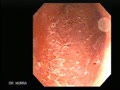
Lesion recognition
- Normal colonic mucosa shows a fine, ramifying vascular pattern.
- Mucosal lesions- The vascular pattern is lost in marked hyperaemia as in IBD.
- There are 9 different endoscopic indices of activity for ulcerative colitis (UC) developed for clinical trials; none have been validated. All 9 indices are subject to interobserver variation (IOV).
- Feagen score for assessing severity of colitis
Stage 1- Granular, hyperaemic mucosa, vascular pattern not visible, not friable
Stage 2- above plus friability (bleeds on contact, but not spontaneously)
Stage 3- above plus spontaneously bleeding
Stage 4- Above plus clear ulceration
Whenever describing a colitis mention at least the extent, whether circumferential or not, friability and presence of ulceration. - The typical endoscopic features of Crohn’s disease are the discontinuos spread of the disease, lesions are distributed asymmetrically. There may be bizarre, ‘map-like’ necroses and fissures. Appearances of so called ‘snail tracks’, aphthoid ulcers etc
- Malignant polyp- is suspected if the polyp is irregular, ulcerated or thick walled. Firmness to palpation with a snare tube is probably the best discriminant for a malignant polyp. If malignancy is suspected, transect low in the stalk and tattoo the area.
- Carcinomas are usually obvious.
- Pseudomembranous colitis- typical membrane like deposits.