Crohn’s disease and diagnosis
Discuss the diagnosis in Crohn’s disease?
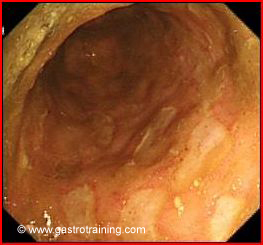
- Colonoscopy is the major tool used to establish the diagnosis of ileocolonic Crohn’s disease. Endoscopic features are focal ulcers and skip areas (segments of normal bowel interrupted by areas of obvious disease)
- Histology is confirmatory. Focal nature of inflammation (as opposed to diffuse pattern in UC) with a relevant clinical history and endoscopy is confirmatory. Granulomas can be seen in up to 30% of patients.
- Wireless capsule endoscopy- another means to visualize the small bowel. It may detect suggestive lesions not visible by other small bowel studies.
- Imaging- It aims to detect the presence of ulceration, fistulas, bowel oedema, strictures, and extraintestinal abnormalities.
- Plain abdominal X-ray- Limited role in assessing bowel inflammation, but can identify bowel obstruction or perforation
- Barium examinations- Barium meal follow through, enteroclysis or barium enema. These tests have a high false negative result. These tests may help in documenting the length and site of strictures or any fistulas.
- Enteroclysis- is performed after placement of a naso-jejunal catheter and infusion of a barium and methylcellulose solution, which provides double contrast images of the small intestine. The technique produces excellent visualisation of the mucosa.
- Fistulography- it is performed to outline fistulous tracks and their communication with the bowel.
- Ultrasound- US of the bowel has high sensitivity for diagnosing inflammatory bowel disease (particularly of the terminal ileum) but is highly operator dependent and comprehensive evaluation of the bowel may be hampered by excessive bowel gas and overlapping bowel loops. Sonographic findings in IBD include increased bowel wall thickening, increased bowel vascularity, and associated mesenteric lymphadenopathy. Reported sensitivity and specificity are 75-94% and 67-100%, respectively.
- CT scan- Computed tomography has high sensitivity for diagnosing intestinal and extraintestinal abnormalities in IBD. However, this technique carries a high radiation dose, which may prohibit its use in young patients or patients who need repeated investigations. Sensitivities and specificities of 73-82% and 70-98%, respectively. Both oral and IV contrast is used. IV contrast may be difficult to use in renal failure patients
- MR enterography- This is performed after oral ingestion of 1.5-2 litres of enteral contrast, which helps to distend the bowel. Sensitivity of 88-98% and specificity of 78-100%. The important advantages of MR compared with computed tomography include better tissue contrast, absence of exposure to radiation, and higher sensitivity for intestinal and extraintestinal changes in Crohn’s disease. The excellent tissue contrast obtained on magnetic resonance imaging can also be used to differentiate between fibrotic and acute inflammatory disease. MR enterography takes 30-40 minutes of scanning time compared to 10-20 seconds with modern CT scanners.
- Blood tests
- CRP- elevated CRP levels correlate with the degree of inflammation.
- Antibody tests — A number of autoantibodies have been detected in patients with IBD some of which may be clinically useful for establishing the diagnosis and differentiating Crohn’s disease from ulcerative colitis.
- Two of the most commonly used antibody tests are antineutrophil cytoplasmic antibodies (pANCA) and anti-Saccharomyces cerevisiae antibodies (ASCA), the combination of which have been proposed as a means for diagnosing IBD and distinguishing Crohn’s disease from ulcerative colitis. pANCA+/ASCA- is suggestive of UC and pANCA-/ASCA+ for CD. The predictive value of antibody tests for the diagnosis of IBD is uncertain at present.
- Stool test- Faecal calprotectin (FC) – It represents 50-60% of neutrophilic cytosolic protein. It is released from cells during cell activation or death. FC is a sensitive marker of bowel inflammation. FC levels correlate well with endoscopic and histological activity in IBD. It is stable in faeces for several days after excretion. It is easily measured in stool by commercially available enzyme-linked immunosorbent assays (ELISA). FC can be used to differentiate flares from IBS like symptoms
- White cell scan- Abdominal scanning with 111-Indium white cell technique visualizes segments of inflamed bowel and quantitates the degree of intestinal inflammatory activity. Its use is on the decline with wider availability of capsule endoscopy and CT and MR enterography.
Pic 1 Colonic ulcers in Crohn’s disease