


Liver
Discuss the liver anatomy?
It is the largest abdominal organ. It is attached to the diaphragm and the anterior abdominal wall by the falciform ligament. The liver is surrounded by Glisson’s capsule of strong connective tissue.
Liver anatomy can be described using two different aspects: morphological anatomy and functional anatomy.
Morphological anatomy is based on external appearances
The falciform ligament on the diaphragmatic surface and left sagittal limb on the visceral surface divides the liver into the right and left anatomic lobes, which are very different from the functional right and left lobes.
On the visceral surface of the liver, there is an H shaped group of fissures and fossa. The crossbar of the H is the porta hepatis. It contains the portal vein, hepatic artery, hepatic ducts, nerves and lymphatics. The left sagittal limb of the H is the fissures containing the ligamentum teres and the ligamentum venosum. The right sagittal limbs of the H are fossae for the gallbladder and IVC.
In this morphological description, the quadrate and caudate lobe belongs to the right lobe of the liver.
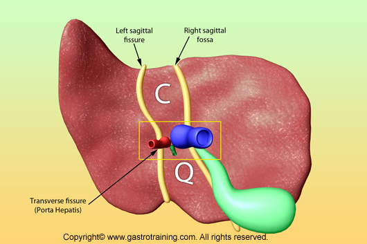
Q= quadrate lobe, C= caudate lobe
Functional anatomy
Functionally the liver is divided into two lobes, right and left by a plane that passes through the gallbladder fossa and the fossa for IVC (i.e. left sagittal limb). Each lobe has its own arterial supply and venous and biliary drainage. Thus the caudate and most of quadrate lobe belong to the functional left lobe of liver.
Within each lobe the branches of portal vein and hepatic artery are consistent and divide each lobe into 4 vascular segments. So the liver is divided into eight functionally independent segments. Each segment has its own vascular inflow, outflow and biliary drainage. In the centre of each segment there is a branch of the portal vein, hepatic artery and bile duct. In the periphery of each segment there is vascular outflow through the hepatic veins.
Because of this division into self-contained units, each segment can be resected without damaging those remaining. For the liver to remain viable, resections must proceed along the vessels that define the peripheries of these segments.
Segment 1 (caudate lobe) is located posteriorly. It is not visible on a frontal view. On a normal frontal view the segments 6 and 7 are not visible because they are located more posteriorly. The right border of the liver is formed by segment 5 and 8. Although segment 4 is part of the left hemi liver, it is situated more to the right.

Right hepatic vein divides the right lobe into anterior and posterior segments
Middle hepatic vein divides the liver into right and left lobes. Left hepatic vein divides the left lobe into a medial (segment 4) and lateral part (segments 2 and 3). The right portal vein divides the right lobe of the liver into superior segments (7 and 8 ) and the inferior segments (5 and 6). During ultrasound it is easy to detect hepatic veins. Hepatic veins are then used to identify vascular segments.
Discuss caudate lobe?
The caudate lobe is often enlarged in cases of cirrhosis and the Budd-Chiari Syndrome. The reason for this is unclear. However, differences in different hormones, nutritional elements, and hepatotropic factors in the portal blood flow between the caudate lobe and the other segment of the liver may correlate with the hyperplastic change of the caudate. Caudate lobe drains directly into IVC (through hepatic veins) and may receive a specific pedicle independent of the portal vein.
Discuss the vascular and lymphatic supply of Liver?
Arterial supply of the liver
Liver has a dual blood supply
Hepatic artery (30%) – it carries oxygenated blood to the liver. The hepatic artery divides into right and left hepatic artery at the porta hepatis
Portal vein (70%) – It carries venous blood containing the products of digestion from the GIT. At the porta hepatis, portal vein terminates by dividing into right and left branches.
Venous drainage of the liver
Hepatic veins drain the liver into the IVC. Hepatic veins are formed by the union of the central veins in its lobules. There are superior (right, left and middle hepatic veins) and inferior group (6-18 small veins draining the right lobe) of hepatic veins.
Lymphatic drainage of the liver
Hepatic nodes along the hepatic vessels (these drain in coeliac nodes)
Some of the lymph vessels follow the hepatic veins to the vena cava foramen in the phrenic and mediastinal nodes.
Discuss the liver histology?
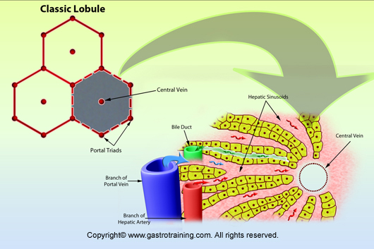
- Hepatic parenchyma can be divided into hexagonal lobules separated by sheets of connective tissue.
- The lobule is the structural unit of the liver, with portal triads (contains branches of portal vein, hepatic artery and bile ductule) at the vertices and a central vein in the middle.
- The parenchymal cells of the liver are hepatocytes. Hepatocytes make contact with blood in sinusoids, lined by highly fenestrated endothelial cells and populated with phagocytic Kupffer cells. The space between endothelium and hepatocytes is called the Space of Disse which collects lymph for delivery to lymphatic capillaries.
- Bile secreted by hepatocytes collect in channels called canaliculi (the basal faces of hepatocytes are joined together by junctional complexes to form canaliculi). These secretions flow toward the periphery of lobules and into bile ductules and interlobular bile ducts, ultimately collecting in the hepatic duct outside the liver.
- Terminal branches of hepatic artery and portal vein empty together and mix in the sinusoids of the liver. Blood flows through the sinusoids and empties into the central vein of each lobule. Central veins coalesce into hepatic veins, which leave the liver and empty into the vena cava.
Pic 1: Liver lobule
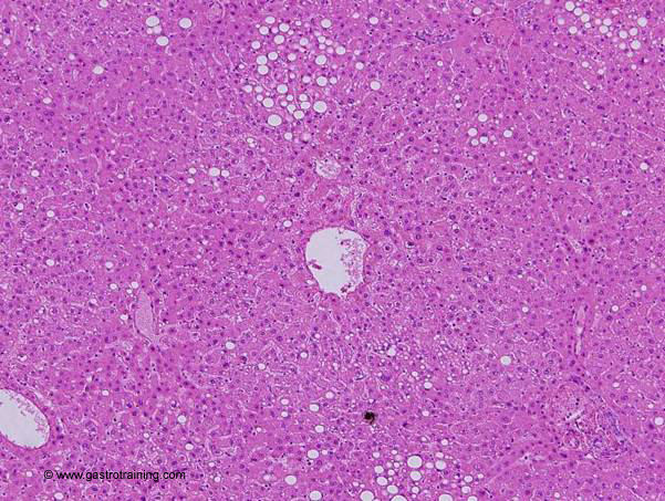
Pic 2: Hepatic lobule -Photo is centred on a lobule with central vein, visible sinusoids, a portal triad to the lower right, some fatty infiltration, and portions of surrounding lobules.
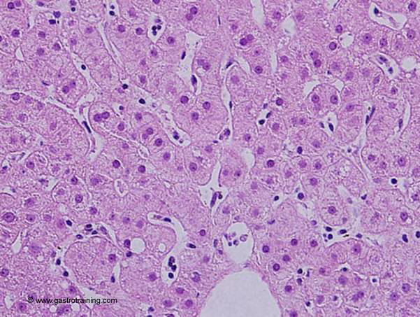
Pic 3: Hepatocytes -note the sinusoids converging on the central vein at the bottom
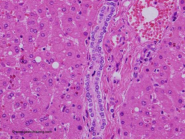
Pic 4: Bile duct -An interlobular bile duct is seen in longitudinal section, with a cross section of a portal vein branch to the right
Ref:

