


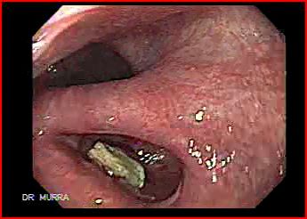
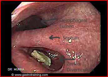
Mr Jones, a 70 year old retired pharmacist presented with complaints of dysphagia and regurgitation of partially digested food material. His endoscopy showed:
What is the diagnosis?
Zenker diverticulum
Discuss the diagnosis of Zenker diverticulum?
Barium swallow is the investigation of choice.
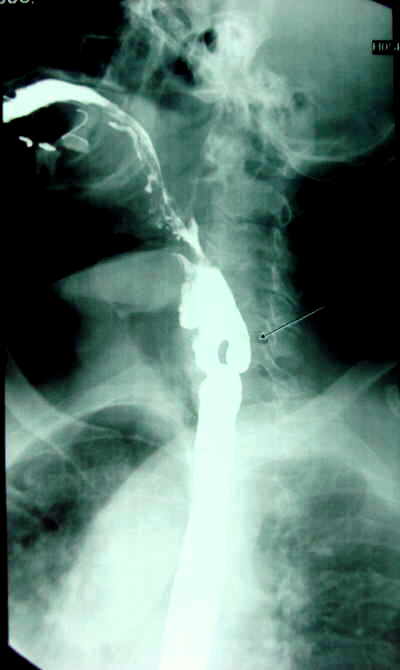
Discuss the role of barium swallow as the first investigation in the diagnosis of dysphagia?
Patients with proximal dysphagia may have a pharyngeal pouch, post cricoid web etc which increases the risk of perforation on endoscopy. Barium swallow may be considered as an initial investigation for investigation of proximal dysphagia; however an endoscopy is safe as an initial test in experienced hands.
What is a Zenker diverticulum?
It is a pulsion diverticulum caused by herniation of the oesophageal mucosa posteriorly between the cricopharyngeus muscle and the inferior pharyngeal constrictor muscles.
It is a false diverticulum consisting of mucosa and submucosa that arises from the posterior portion of the inferior pharyngeal constrictor muscle. True diverticulum consists of all layers of the wall.
Zenker diverticulum occurs at an area of potential weakness in the inferior pharyngeal constrictor muscle referred to as the Killian dehiscence. It is located between the oblique fibers of the inferior constrictor muscle and the horizontal fibers of the cricopharyngeal muscle.
The aetiology is incompletely understood. It is hypothesized that improperly timed relaxation of the cricopharyngeus muscle during swallowing leads to increased pressure which causes herniation of the oesophageal mucosa over time.
What are the symptoms of Zenker diverticulum?
It affects older patients (males slightly more than females)
Dysphagia is the commonest symptom occurring in as many as 98% of patients.
Other common symptoms include halitosis, regurgitation of undigested food, noisy swallowing, and aspiration. Hoarseness can be present when the diverticulum is large enough to compress the recurrent laryngeal nerve.
Weight loss and recurrent pulmonary infections secondary to aspiration occur in approximately one third of patients.
Discuss the treatment of Zenker diverticulum?
Zenker diverticula require intervention only if they produce symptoms. In general, small (i.e., <2 cm) lesions found incidentally require no intervention.
The two key elements of the successful surgical management of Zenker diverticulum are division of the cricopharyngeus muscle to eliminate the potentially elevated pressure zone and elimination of the diverticular pouch as a reservoir of food and secretions.
Treatment options:
Surgical
Small Zenker diverticulum- Cricopharyngeal myotomy (muscle is divided longitudinally) via a left neck incision plus diverticulopexy (diverticulum is inverted and sutured to the prevertebral fascia i.e. diverticulum is not excided).
Larger Zenker diverticulum- Cricopharyngeal myotomy plus diverticulectomy (pouch neck is stapled and then the pouch is excised)
Endoscopic
Endoscopic approach has become the preferred choice for most American and British surgeons. This technique consists of adhering the mucosal edges by stapling them together while cutting across the “party wall” i.e. septum at the same time. Few complications have been reported with this procedure.
Alternatively, flexible endoscopes are used nowadays to cut (using APC or needle knife) the septum between the diverticulum and the oesophageal lumen. The septum contains the cricopharyngeal muscle.
Image courtesy of www.gastrointestinalatlas.com

