


Mrs Bennett a 65 year woman presents with dysphagia. Endoscopy showed:
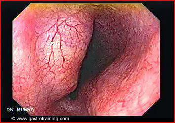
What is the diagnosis?
Oesophageal subepithelial lesion
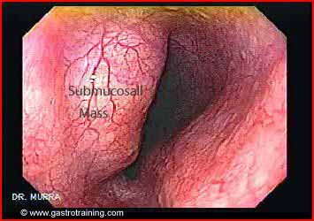
Discuss the differential diagnosis of an oesophageal subepithelial lesion?
- Leiomyoma- is the commonest cause of subepithelial lesions in oesophagus. These are benign mesenchymal tumours of the GI tract. They usually occur in the distal third of the oesophagus.
- GIST- are mesenchymal tumours originating from the interstitial cells of Cajal (ICCs) inside the muscle layer
- Lipomas- are benign tumours of adipose tissue arising from the submucosa. Endoscopically, they are usually seen as a smooth bulge with a yellowish hue and normal overlying mucosa. A ‘pillow sign’ can be detected by pushing the biopsy forceps into the soft tumor, leaving an indentation.
- Duplication cysts- are congenital abnormalities that develop as the result of an error in recanalization of the embryonic gastrointestinal tract. Duplication cysts may be contained within the wall of the GI tract or may be extrinsic, often closely adhered to the wall of the gut.
Discuss the role of biopsy in oesophageal subepithelial lesions?
Endoscopic biopsies are unhelpful in diagnosing the aetiology of a subepithelial lesion.
Discuss the diagnostic work up of an oesophageal subepithelial lesion?
Endoscopic ultrasound is the investigation of choice in diagnosing the aetiology of an oesophageal submucosal lesion. Further EUS guided FNA helps in establishing a histologic diagnosis.
CT scan may be an alternative in units where EUS is not available. It may help if the lesion is large; however it is not as sensitive as EUS.
Discuss EUS for subepithelial lesions of GIT?
EUS plays a pivotal role in the description, diagnosis, and management of these lesions. EUS can distinguish intramural lesions from extrinsic compression, and can often accurately diagnose the nature of these lesions by their echo characteristics and wall layer of origin. Thus GIST and leiomyomas arise from the muscle layer and are hypoechoic. The lipomas arise from the submucosa and are bright or echogenic.
Discuss the EUS characteristics of oesophageal subepithelial lesions?
Leiomyomas are generally more homogeneous lesions and lacks findings of cystic spaces and irregular borders on EUS examination, which are features more often found in GISTs. However, leiomyomas can sometimes be indistinguishable from GISTs. Therefore, tissue diagnosis should be sought
Lipomas appear as hyperechoic, homogeneous lesions arising from the submucosa. The diagnosis can often be made by their endoscopic appearance alone. In those lesions where the diagnosis is uncertain, EUS can be helpful. Because of their classic, characteristic EUS appearance, fine-needle aspiration is not necessary for diagnosis.
Duplication cysts- appear as round, anechoic lesions arising from the oesophageal wall or may be extrinsic to the gastrointestinal wall. These cysts may contain thick mucinous material, septations, fluid levels, or debris. EUS characteristics alone are usually sufficient for diagnosis. If the diagnosis is in question or there is suspicion of malignancy, fine-needle aspiration may be done.
Discuss the clinical manifestations of oesophageal subepithelial lesions?
The majority of subepithelial tumors are asymptomatic and often are found incidentally during endoscopy or radiologic examinations performed for other reasons. Large lesions may cause dysphagia.
Discuss the management?
Surgical resection is recommended in cases of symptomatic leiomyomas or lipomas. Asymptomatic tumors do not require any intervention or endoscopic follow-up.
Management of GIST
Duplication cysts- are usually found incidentally on endoscopy or radiographic imaging. There is no consensus regarding the management of asymptomatic duplication cysts. Management options range from performing surgical resection to prevent future complications (dysphagia, haemorrhage, perforation etc) to periodic follow-up with EUS. Symptomatic cysts can be treated surgically with resection or marsupialization. They can also be treated successfully endoscopically using fine-needle aspiration.
Discuss the layers of GI tract seen on EUS?
- EUS provides high resolution imaging of the GI tract by producing an image of the GI tract wall consisting of five sections or layers. These layers correspond to the five distinct histologic layers of the gut wall:
- The innermost layer (lumen) is hyperechoic or echogenic, and is attributed to the initial echo-interface between the ultrasound waves, the GI tract mucosa, and surrounding fluid.
- The second layer is a hypoechoic or dark band that corresponds to the actual mucosa
- The third layer is a hyperechoic or bright echo that corresponds to the submucosa.
- The fourth layer represents the muscularis propria and is depicted as a hypoechoic.
- The fifth layer, which is seen as a hyperechoic or bright echogenic band, is the serosa in the stomach, duodenum, and rectum and the adventitia in the oesophagus.
Further reading
Link to GIST
Image courtesy of www.gastrointestinalatlas.com

