Peptic ulcer disease (PUD)
Discuss peptic ulcers?
Peptic ulcers are defects in the gastrointestinal mucosa that extend through the muscularis mucosae. The two main causes of PUD are HP infection and NSAIDS. However about 15% of peptic ulcers are idiopathic or due to rare causes (rare causes -drugs like Sirolimus, gastrinoma, systemic mastocytosis, carcinoid syndrome, ischaemia, radiation injury, sarcoidosis, Crohn’s disease etc). In addition COPD and cirrhosis is associated with an increased risk of PUD. There is no evidence that alcohol is a risk factor for peptic ulcer. Further steroids alone are not associated with an increase in the risk for peptic ulcer. However, steroids may worsen NSAID-induced ulceration.
What are the Clinical manifestations of peptic ulcer disease?
Epigastric discomfort- Classic pain of duodenal ulcer occurs in empty stomach whereas gastric ulcer (GU) causes more severe pain occurring soon after meals.
Discuss diagnosis of PUD?
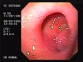
PUD is very often diagnosed at endoscopy. Benign ulcers have smooth, regular and rounded edges. The following features are suggestive of malignancy on endoscopy:
- The gastric folds around the ulcer are nodular, fused, or stop short of the ulcer margin
- The ulcers margins are irregular or thickened
Discuss treatment of peptic ulcer disease?
H. Pylori positive ulcers- Eradicate H. Pylori. Anti secretary therapy for 4-12 weeks after eradication treatment may be appropriate, especially in patients with a complicated peptic ulcer. Prolonged anti secretory therapy can be justified in patients who are considered to be at high risk, since no studies have had the power to define the optimal management in these patients.
H. pylori negative ulcers —Anti secretory drugs for 4-6 weeks. At least two different gold standard tests should be negative before a confident diagnosis of H. pylori infection-negative peptic ulcer disease can be made.
Discuss refractory ulcers?
Ulcers are considered refractory if healing is not evident after 8 to 12 weeks of therapy.
Causes of refractory ulcers- persistent H. pylori infection, continuing NSAID use, cigarette smoking (contribute to impaired healing), giant ulcers (take longer to heal) and poor compliance. Occasionally refractory cases are due to gastrinoma or ulcer cancer.
Serum fasting gastrin level may be needed as a screening test for Zollinger-Ellison syndrome. Surgery should be considered if the above causes of refractory ulcer are excluded.
Discuss recurrent ulcers?
H. pylori ulcers recur within six months in up to 20 percent of patients after apparently successful antibiotic treatment. Other factors are failed eradication of H. pylori, gastrinoma, and surreptious use of NSAIDs.
Recurrent ulcers patients are more likely to have persisting acid hypersecretion. Thus, patients with recurrent ulcers should be maintained on anti secretory therapy.
Discuss endoscopic follow-up after peptic ulcer treatment?
Duodenal ulcers — Patients with uncomplicated DU do not need further endoscopy unless symptoms persist or recur. HP eradication should be confirmed.
Gastric ulcers- Repeat endoscopy to confirm GU healing is the standard practice to ensure that the lesions are benign. However, a case can be made against the practice, if the initial biopsies are adequate (at least four good biopsies from the ulcer margin and one from the base).
Discuss the pharmacotherapy in bleeding peptic ulcer?
Patients with high risk stigmata (such as a visible vessel or adherent clots) are at high risk of rebleeding. 80 mg bolus (of omeprazole or pantoprazole) followed by 8 mg/hr infusion has become the standard in clinical practice after endoscopic treatment of the ulcer. If there is no rebleeding within 24 hours, the patient may be switched to oral pantoprazole 40 mg/day or omeprazole 20 mg/day. Twice daily dosing of an oral or IV proton pump inhibitor may also be a reasonable alternative.
Somatostatin and octreotide — Somatostatin or its long-acting analogue octreotide have a theoretical benefit in bleeding ulcer disease because they reduce splanchnic blood flow, inhibit gastric acid secretion, and may have gastric cytoprotective effects. A meta-analysis of trials suggested that somatostatin was associated with a reduced risk of continued bleeding (relative risk 0.53 (95 percent CI, 0.43 to 0.63)). The risk also appeared to be reduced by octreotide, although there were fewer studies. Thus, somatostatin or octreotide can be used as adjunctive therapy before endoscopy, or when endoscopy is unsuccessful, contraindicated, or unavailable. A typical dose of somatostatin was 250 micrograms given as a bolus then given hourly for three to seven days, while a typical dose of octreotide was 50 to 100 micrograms given as a bolus followed by 25 micrograms/hour for up to three days.
Discuss complications of peptic ulcer disease?
There are four major complications of peptic ulcer: bleeding, perforation, penetration and gastric outlet obstruction.
Bleeding peptic ulcer- surgery may be required
Duodenal ulcer — the first priority during emergency surgery is control of the bleeding site. Control is obtained by suture ligature and/or by ligating the gastroduodenal artery at the superior and inferior aspect of the ulcer. Once the bleeding has been addressed, a definitive acid-reducing operation may be performed.
Gastric ulcer — Resection with Billroth I or II reconstruction is generally indicated for bleeding gastric ulcers because of the risk of malignancy. Excision alone is associated with rebleeding in as many as 20 percent of patients so that distal gastrectomy is generally preferred, although excision combined with vagotomy and pyloroplasty may be considered in high risk patients.
Perforation- The presence of free air suggests perforation, although about 10-20 percent of patients with a perforated DU will not have free air.
Simple patch closure or truncal vagotomy with pyloroplasty (incorporating the perforation) is the traditional procedure for perforated duodenal ulcers.
Distal gastrectomy is the preferred surgery in perforated gastric ulcer. Patch closure alone is associated with postoperative gastric obstruction in approximately 15 percent of cases. Laparoscopic repair appears to be a reasonable option.
Penetration- Ulcer penetration refers to penetration of the ulcer through the bowel wall without free perforation and leakage of luminal contents into the peritoneal cavity. Penetration can occur into the pancreas, biliary tract, liver, greater omentum, colon, or vascular structures.
Penetration often comes to attention because of a change in symptoms or involvement of adjacent structures. The change in symptom pattern involves a loss of cyclicity of the pain with meals, and loss of food and antacid relief. The pain becomes more intense and lasts longer. The diagnosis of penetrating ulcer is suspected clinically when an ulcer in the proper region is found.
Gastric outlet obstruction- is the least frequent ulcer complication. Initial treatment includes nasogastric suction, IV fluids to correct fluid and electrolyte deficits and IV PPI. Approximately 50% of cases of gastric outlet obstruction due to peptic ulcer respond initially to this regimen, although some initial responders may eventually require surgery or endoscopic dilatation. All cases require endoscopy and biopsies to exclude malignancy, diagnose ulcer disease, and determine if H. pylori is present.
Endoscopic balloon dilatation is useful for patients who do not respond to initial medical therapy, although repeated sessions are necessary in some cases. Dilatation can be accomplished by using endoscopy and a TTS (through the scope) balloon (sustained for 1 min and 3-4 times), or by using a balloon placed over a guidewire positioned under fluoroscopic guidance. Symptoms are usually considerably improved with successful dilatation to 12 mm. There may be an advantage to postponing dilatation beyond 15 mm until after a period medical management. In one study, for example, 24 of 30 patients (80 percent) achieved sustained symptom relief; 17 had a single procedure while seven required multiple sessions. Dilation failed in four patients with long duodenal strictures, while two dilated to 18 mm suffered perforation; both recovered uneventfully after surgery. A good long-term response is achieved when H. pylori can be cured or NSAID use discontinued.
There is a risk of perforation and hence patients should be appropriately prepared for surgery before dilatation, and monitored closely after dilatation before resuming oral intake. An immediate post-procedure Gastrograffin study is an appropriate precaution to detect perforation in a timely fashion.
Surgical therapy — if the pyloric channel can be identified at endoscopy and a balloon passed, dilatation is an appropriate option in experienced hands. Cases that clearly warrant surgery are ones where the pylorus is obstructed and cannot be safely dilated, or where the obstruction persists or recurs during medical management.
When surgery is necessary, inflammation and scarring may prevent safe antrectomy, which would otherwise be an excellent choice since it resects the ulcer and relieves the obstruction. In these instances, truncal vagotomy and gastrojejunostomy may be the preferred approach.
Discuss the pathogenesis of NSAIDs induced PUD?
PUD secondary to NSAIDs is due to systemic (post-absorptive) inhibition of GI mucosal cyclo oxygenase (COX-1) activity. So IV or IM NSAIDS can also cause PUD. COX-1 inhibition reduces mucosal production of protective prostaglandins like PGE2.
Mucus and bicarbonate secretion forms a protective layer on the surface of the gastric mucosa, which retards diffusion of acid-pepsin from the lumen into the mucosa. These protective functions are compromised by COX-1 inhibition leading to peptic ulcer formation.
What are the strategies to avoid NSAID induced PUD?
- Using selective COX-2 inhibitors (risk is reduced as none of COX 2 is completely selective).
- COX-2 inhibitors are associated with a modest reduction in the risk of GI bleed compared with nonselective NSAIDs.
- Co-prescribing either a prostaglandin E analog such as misoprostol (200 µg four times daily) or a PPI (Lansoprazole 15 or 30mg daily or esomeprazole 20 or 40 mg daily)
- Enteric coated is designed to resist disintegration in stomach. It diminishes the endoscopic signs of gastroduodenal injury, however it does not reduce the risk of GI bleed. This is not surprising, since injury severe enough to induce bleeding is thought to reflect the systemic rather than the local effects of aspirin.
- H. Pylori eradication- may be considered prior to NSAID use in patients with a past h/o PUD. HP eradication may also be considered prior to considering prior to long term NSAIDs treatment.
The American College of Gastroenterology has identified the five most important variables that place patients at risk for NSAID-related gastrointestinal complications:
- Prior history of a gastrointestinal event (ulcer, hemorrhage)
- Age >60
- High dosage of a NSAID
- Concurrent use of glucocorticoids
- Concurrent use of anticoagulants
Patients with these risk factors should be considered for prophylaxis with NSAID treatment
Pic 1 Duodenal ulcer